Another weekend shift and another emergency case.!!!
This lady recently came into the hospital at 30 weeks pregnant. The clinical
details I received were ? Pre-eclampsia.
The Full Blood Count
showed that the platelet count for this lady was low at 64 x 10^9/l and had significantly dropped
from 266 x 10^9/l, two weeks previously. This situation must always
be dealt with immediately. The first question is, is this a genuine result?
After ruling out a clot in the sample, or platelet
clumping/ Fibrin strands, the next question is what is going on in the body to
make the platelets fall like this? The Scientist must then seek the answer by
looking at the white cells and red cells on the blood film, clinical details
and other laboratory results.
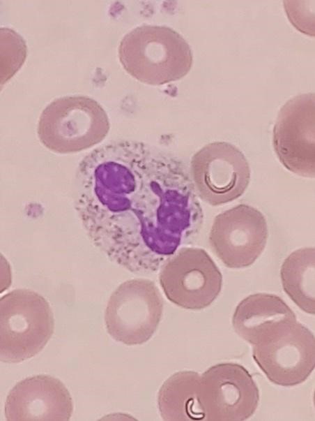
The potential cause was revealed on the blood film, by the
presence of red cell fragments in most fields. This is a serious finding in conjunction with a dropping platelet count and the question is, why are
red cells being sheared in half!?

My immediate thought was HELLP Syndrome which stands for Haemolysis, Elevated Liver Enzymes
and Low Platelets. This is a severe, potentially life -threatening form of
pre-eclampsia. Complications include liver haemorrhage or rupture, pulmonary
odema, placental abruption, bleeding and clotting issues.
What is the cause for
red cells being sheared in half ?
It seems that the main cause is a Microangiopathic
Haemolytic anaemia (MAHA). The red cells are sheared off as they pass through
capillaries with damaged endothelium and fibrin strands which leads to the red
cells being fragmented as they pass through.
Another cause for the red cell damage can also be
Disseminated Intravascular Coagulation (DIC).
Why are platelets
reduced in HELLP?
The platelets are aggregating and forming clots due to
endothelial damage.
Further evidence
that this was HELLP Syndrome.
Protein in the urine
A protein: creatinine
ratio or >30mg/mmol suggests significant proteinuria in pregnancy (NICE, 2019). In this case the value was
341.7mg/mmol!
Elevated Liver
enzymes
Nice guidelines ( NICE, 2019) suggest a rise in ALT, twice
the upper limit of the normal range is of concern. The ALT in this case on
presentation was 420 U/L. The normal range is <33 U/L.
What happened next?
The obstetric team and Consultant Haematologist were alerted
to the blood film findings and other laboratory results. The Consultant
Haematologist should be informed as red cell fragments with low platelets could
also be suggestive or other life threatening microangiopathic haemolytic anaemias such as
TTP and HUS, where the course of treatment would be entirely different.
The diagnosis of HELLP was indeed made in this case however and the
decision to deliver the baby prematurely, despite the lady only being 30 weeks pregnant. Delivery
is the cornerstone of treatment for HELLP syndrome (Baha, 2022).
Post delivery we can see quite a quick improvement with an upward trend in the platelet count and downward trend in
ALT. From a haematological point of view,
if the platelet count did not improve, an alternate cause for the
thrombocytopenia such as a primary Microangiopathic Haemolytic anaemia would be
sort.
|
|
Haemoglobin (g/dl)
|
Platelets (x 109/l)
|
ALT (U/L)
|
|
PRESENTATION |
118
|
64
|
420
|
|
DAY 1 (post delivery)
|
111
|
114
|
300
|
|
DAY 2
|
98
|
178
|
212
|
|
DAY 3
|
99
|
239
|
117
|
|
DAY 4
|
102
|
337
|
85
|
|
DAY 8
|
108
|
558
|
32
|
|
DAY 15
|
119
|
354
|
14
|
Hopefuly mother and baby both had positive outcomes in this
case.
References
Baha, S., 2022. UpToDate.
[online] Uptodate.com. Available at:
<https://www.uptodate.com/contents/hellp-syndrome-hemolysis-elevated-liver-enzymes-and-low-platelets?search=hellp%20SYNDROME§ionRank=3&usage_type=default&anchor=H24&source=machineLearnin>
[Accessed 8 May 2022].
Petca, A., Miron, B., Pacu,
I., Dumitrașcu, M., Mehedințu, C., Șandru, F., Petca, R. and Rotar, I., 2022.
HELLP Syndrome—Holistic Insight into Pathophysiology. Medicina, 58(2),
p.326.
Nice.org.uk.
2022. Recommendations | Hypertension in
pregnancy: diagnosis and management | Guidance | NICE. [online] Available
at:
<https://www.nice.org.uk/guidance/ng133/chapter/Recommendations#assessment-of-proteinuria-in-hypertensive-disorders-of-pregnancy>
[Accessed 8 May 2022].