About Me

My photo
I'm a busy Mum and a Biomedical Scientist in Haematology. My particular interest is in blood cell morphology and parasitology, where I never stop learning.

Saturday 19 February 2022

blood film diagnosis- it's all about texture, colour and shape!


Recognising an acute leukaemia heavily depends on your ability to make the distinction between different textures, colours and shapes!

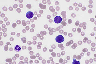
Take a look at this photo of a blast cell (upper left) seen in an Acute Myeloid Leukaemia and a mature neutrophil (cell that looks like a happy face, bottom right). Notice the nucleus. The blast cell has chromatin in the nucleus that is very spread out, almost looks as if dots have been made with a fine pen. Compare this to the neutrophil. The chromatin in the nucleus have condensed, showing lots of texture. To me it almost looks like knots!


Blasts often have spherical inclusion in the nucleus called a nucleolus, which can be seen on the cell above over to the left side. This also tells us we are looking at an immature cell.

Scientists use this along with features such as size of cell and size of nucleus compared to cytoplasm,  in order to recognise an immature cell. 

From there we need to take this further and distinguish which type of Acute Leukaemia we could be dealing with and again this about recognising subtle features. Look at this picture. The little red ‘comma’ like inclusion in the cytoplasm is called an auer rod. This tells us that this is an Acute Myeloid as opposed to an Acute Lymphoid Leukaemia.


To go further it is possible morphologically to determine more about the type of Acute Myeloid Leukaemia we are dealing with, by looking at shapes and colours and this leads me onto today’s film which my colleague very kindly saved for me . It’s an Acute Promyelocytic Leukaemia (APL). It is vital that the distinctive morphology is not missed as APL is a haematological emergency.

 There are two types of APL, one has abnormal promyelocytes with numerous granules, whilst the other which we have here is the variant form and the blasts have distinctive bilobed nuclei and appear to lack granules. The morphology of the variant is quite distinctive and should always be on the Scientist's mind when assessing a blood film for a new leukaemia.


APL is a haematological emergency as it can be rapidly fatal due to bleeding and thrombotic complications. This patient indeed had a D-Dimer result of 2560 ng/ml. Suggestive of DIC. In DIC clotting factors are abnormally consumed which leads to not only bleeding but also thrombotic issues. Bleeding into the brain or lungs or clots blocking the heart, brain and  lungs can be life threatening.

I’ve been reading an article that explains APL quite nicely at https://rarediseases.org/rare-diseases/acute-promyelocytic-leukemia.

I didn’t know the actual reason behind the coagulation issues in APL but it appears that on the surface of the abnormal promyelocytes is tissue factor which contributes to the activation of the clotting cascade. Also Annexin II, present on the surface of the promyelocytes activates plasmin which breaks down blood clots. The activation of these two leads to excessive clotting, excessive bleeding and excessive breaking down of clots.

The treatment for this type of leukaemia uses a derivative of vitamin A which matures the promyelocytes to mature neutrophils! This treatment is called al-trans-retinoic acid or ATRA.  This is because most patient’s with APL, alterations in the RARA gene occur, which is involved in using vitamin A to mature blood cells. The alteration to the gene occurs where the RARA and PML genes are located leading to the PML/RARA fusion gene. This leads to abnormal vitamin A and stops maturation of the promyelocytes, leaving them in effect ..stuck! Pretty clever treatment based on understanding the role of the RARA gene.

The ability to recognise differences in texture, colour, shape and size of blood cells forms the basis of blood cell morphology . This is vital in the rapid diagnosis and treatment of APL because quick treatment with a vitamin A derivative is required to mature the abnormal promyelocytes and therefore lessen the coagulopathy.


Tuesday 15 February 2022

Antithrombin


I asked a colleague who is very experienced in thrombophilia testing and interpretation, how I could improve my…er.....not so good knowledge! She suggested Practical-Haemostasis.com. It’s a free website and I’ve found it really useful.

So my task tonight is Antithrombin.

Antithrombin (AT) s a natural anticoagulant and has a very important role in stopping the action of Thrombin ( IIa), Factor Xa (FXa) and to a lesser extent XIa (FXIa) and IXa (FIXa). These are called Serine Proteases which means they cut peptide bonds in specific proteins. So basically AT is involved in preventing the body from clotting to much which can lead to thrombosis. If a patient is deficient in this, Venous Thromboembolic disease (VTE) can occur.

I didn’t realise that there are six types of AT. The most significant was known as Antithrombin III, but the III has been dropped and is now known simply as antithrombin.

The laboratory I work in measures this by a chromogenic method, so in short by a colour change, which allows the absorbance (light at a particular wavelength) to pass through a volume of liquid. Plasma is incubated with an excess of the relevant substrate. In my labs case it is with an excess of FXa in the presence of excess heparin.

The heparin changes the structure of the AT to increase it’s activity as an inhibitor. A chromogenic substrate is the added which is specific for the enzyme FXa. Any residual cause the substrate to be cleaved and a colour change. The absorbance at 405nm is inversely proportional to the amount of antithrombin activity in the plasma.

AT can also be measured immunologically.

Result interpretation should always bear in mind the limitations of a particular method. The bovine FIIa method is suggested to be the best at detecting all AT variants. The human FIIa and bovine FXa may not detect all clinically significant variants.

So it seems that AT deficiency can be inherited or acquired. Acquired causes can be seen in liver dysfunction, Sepsis, DIC, Pre-eclampsia, AML, Heparin therapy, Proteinuria, in association with L-Asparaginase,, Chron’s Ulcerative Colitis, Poor nutrition and dilutional reasons such as haemodialysis , Cardiopulmonary bypass and Plasmaphoresis.

I’ve been reading my Trust’s guidance for thrombophilia testing and acquired AT deficiency is not tested for. Inherited deficiencies are investigated if diagnosis will affect patient management. So for example patients who will be on long term anticoagulants due to recurrent VTE and unprovoked DVT or PE. Also decisions regarding thrombophylaxis in pregnancy. Unusual sites for thrombosis and arterial thrombosis may also warrant thrombophillua screening.

Thrombophillia testing is not recommended in decisions regarding contraception using oestrogen but instead a progesterone only preparation is recommended. 

In pregnancy morbidity, it is suggested that just Antiphospholipid antibodies are tested on two separate occasions at least six weeks apart. Whilst patients developing warfarin skin necrosis,  a Protein C and Protein S are indicated.

And I'll leave today's learning there and move on next time to another part of thrombophillia screening.