About Me

My photo
I'm a busy Mum and a Biomedical Scientist in Haematology. My particular interest is in blood cell morphology and parasitology, where I never stop learning.

Friday 17 May 2024

Proposal for Percentage Schistocyte Counts in patients with a low or dropping platelet count with schistocytes on blood film.


                                                                   Zini et al, 2012


 I recently reviewed the ICSH recommendations for counting schistocytes in a suspected Thrombotic Microangioapathy and have proposed we do this in our laboratory.

Proposal

To provide a percentage schistocyte count on those films where there is a true thrombocytopenia in conjunction with schistocytes.

The ICSH gives normal reference value of <1 % schistocytes in healthy adults and full term neonates and <5% in premature neonates2 .It is suggested that schistocyte counts greater than these values are a robust morphological threshold for suspecting red cell mechanical damage due to thrombotic microangiopathy (TMA). A lack of schistocytes does not rule out a TMA however.

When not to perform a schistocyte count

Schistocytes are seen in a number of conditions and are not specific to TMA. Schistocytes caused by a TMA are usually the dominant feature on the blood film, perhaps in conjunction with moderate signs of stimulated erythropoiesis such as polychromasia, basophilic stippling and nucleated red cells.

If schistocytes are present alongside multiple other red cell abnormalities, the percentage schistocyte count is not appropriate.

Criteria for schistocyte recognition

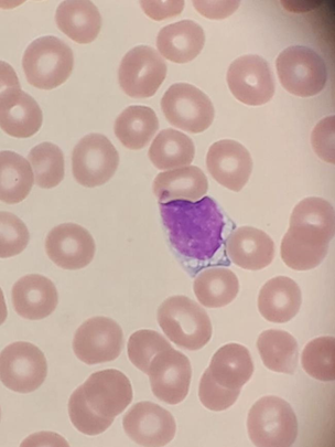
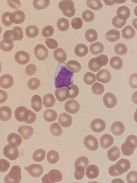
Schistocytes are defined by the ICSH as always smaller than intact red cells and can have the shape of fragments with sharp angles and straight borders, small crescents, helmet cells, keratocytes, or microspherocytes. Microspherocytes only to be included in the presence of other shapes mentioned1

Bite cells, spherocytes, irregularly contracted cells, tear drop poikilocytes, and echinocytes should not be included in a schistocyte count.

(See photograph above)

Method

Quantify the schistocytes, only if it they are the dominant abnormality on the film. Not appropriate if they are present alongside multiple other red cell abnormalities.

·         Schistocytes will be counted using a high power x 100 objective.

·         Work out by counting, approximately how many red cells are present per field On a well spread normal film this is approximately 200.

·         In each field count how many schistocytes you see until you have covered 1000 red cells.

·         Express as a percentage of red cells.

Example

Worked out that there are approximately 200 red cells per high power field. Need to count how many fragments are in 10000 red cells. Therefore, five fields of 200 cells will give a total of 10000 red cells.


Field 1   200 red cells, 10 schistocytes seen

Field 2   200 red cells, 6 schistocytes seen

Field 3   200 red cells, 8 schistocytes seen

Field 4   200 red cells, 8 schistocytes seen

Field 5   200 red cells, 9 schistocytes seen


10+6+8+8+9 = 41 schistocytes seen in a 10000 red cells


41  10000 x 100 = a schistocyte count of 4.1%


Schistocyte recognition.


a) Keratocyte and helmet cell, b) Schistocyte, c)microspherocyte

Zini et al, 2021

References

1 Zini G, d'Onofrio G, Biggs C, et al. ICSH recommendations for identification, diagnostic value, and quantitation of schistocytes. Int J Lab Hematol. 2012; 34: 107-116.

2 Zini G, d'Onofrio G, Erber WN, Lee SH, Nagai Y, Basak GW, Lesesve JF; International Council for Standardization in Hematology (ICSH). 2021 update of the 2012 ICSH Recommendations for identification, diagnostic value, and quantitation of schistocytes: Impact and revisions. Int J Lab Hematol. 2021 Dec;43(6):1264-1271. doi: 10.1111/ijlh.13682. Epub 2021 Aug 24. PMID: 34431220.

Monday 20 November 2023

Which Parameters are truly useful in XbarM analysis and which are a waste of time!

Reviewing the XbarM analysis parameters used in our laboratory XbarM control program and looking at whether the current parameters being monitored are providing truly useful information on anlayser and reagent performance. I read this paper https://www.sysmex.co.uk/education/library/documents/detail/the-xbarm-control-program-use-it-wisely.html, which is really helpful. The section I found particularly interesting is where parameters are divided into three groups depending on their significance in analyser monitoring. These groups should be observed and dealt with differently as some have very little biological variation and are not effected by the patient population whereas others depend on a multitude of variables. These are named ‘major’, ‘supporting’ and ‘other’ parameters.
Major parameters- These are useful! 
The paper states that the major parameters are mostly independent of factors such as gender and age and patient population and therefore are very useful in XBarM monitoring. These include the MCHC and Sensitivity parameters. They also are involved in two technical units of the XN analysers, the FCM detector and RBC/platelet detector and HGB detector.
MCHC
The MCHC only varies naturally within a tight range as it is calculated from the HGB and HCT and abnormal results for the MCHC are usually related to sample quality issues or analyser failure, rather than clinical reasons. The clinical reasons I can think of are slightly raised MCHC can be seen when spherocytes are present in haemolysis and low value can been seen when there is a microcytic, hypochromic anaemia. On the XN series, the MCHC is two different technical detectors, the RBC/PLT channel and haemoglobin channel are used and can therefore be monitored at the same time.
 
Sensitivity Parameters 
The sensitivity parameters again are useful in XbarM analysis as biologically will not vary very much and will monitor the technical units used for flow cytometry . They relate to the scattergrams that are produced in different measurement channels and the position of the cell populations. For example below the WDF-X and WDF-Y will mark the position of the neutrophil population on the WDF scattergram and this shouldn’t vary significantly biologically.
Supporting Parameters- Less useful This group is not so important in XbarM analysis as they do have more biological variation and do depend on the patient population . IQC is a better way of monitoring these. These include MCH and MCV, WBC, RBC, PLTS, HGB and HCT. 

Other parameters- Waste of time!!! These again are highly variable parameters that depend on patient population, type of ward, season etc. These are therefore not useful in XbarM control program. They mainly include the WBC differential and distribution parameters such as RDW for example. 

Those parameters that have a very narrow biological range i.e MCHC and sensitivity parameters are very useful in monitoring analyser performance including reagents used, stability of analyser performance and sensitivity of the electronics. For the supporting parameter and other parameters, it would be far better to monitor these by IQC.

Tuesday 26 September 2023

Unstable Haemoglobin Causing Inaccurate White Cell Differential on Sysmex XN


Look at these white cell differentials analysed on the Sysmex XN. The first run to the right labelled  run 1, gives a neutrophil count of 1.21 x 10⁹/L and Eosinophils of 8.5 x 10⁹/l. The white cell differential on second run is completely reversed.


This can also be clearly seen on the WDF plots. On run 1, the large orange population of white cells were counted as eosinophils.  On the second run, they were counted as neutrophils represented by the blue population.


Run 1


Run 2


It is also clear that the white cell populations are ‘squashed’ and restricted to lower portion of the plot. Usually all populations would be higher up the plot, with the monocytes being the highest . This is because side flouorescent light (SFL) corresponds to the amount of DNA and RNA in a cell and monocytes have more than other normal white cells.

The inaccuracy of the white cell differential here, was caused by an unstable haemoglobin. These are haemoglobinopathies where the solubility of haemoglobin is altered, leading to precipitates of haemoglobin and haemolytic anaemia due to reduced red cell lifespan.

The reason the unstable haemoglobin leads to inaccuracies in the white cell differential, is because it does not act in the same way as it’s stable counterparts when the red cells are lysed with Lysercell WDF on the Sysmex XNs.. This is an important step in the measurement of white blood cells. Reading up on this White_Paper_RBC_Diseases.pdf (sysmex-europe.com), it is suggested that the unstable haemoglobin interferes with the fluorescence marker used to differentiate white blood cell populations, leading to decreased flouorescence and inaccurate differentials. This is why the entire plot is shifted down and appears that all cells have little DNA or RNA content.

Therefore if you encounter an abnormal plot where the fluorescence is reduced, it is firstly important to perform a manual differential. Secondly there should be the suspicion of an unstable haemoglobin particularly if haemolysis is present and HPLC and or/genetic sequencing would be indicated.


Tuesday 25 October 2022

Gap analysis on BCSH 2022 Guidelines on the Laboratory Diagnosis of Malaria

 Today I used the BCSH audit template to perform a gap analysis on our procedures for the diagnosis of malaria,  in comparison to the BCSH 2022 guidelines. British Society for Haematology guidelines for the laboratory diagnosis of malaria - Rogers - 2022 - British Journal of Haematology - Wiley Online Library.

 I would recommend that all staff involved in laboratory malaria diagnosis, read the 2022 guidelines and also the following:

WHO information on ‘False Negative RDT results and p.falciparum histidine-rich protein 2/3 gene deletions MAY 2016 (REV. SEPTEMBER 2017 AND JULY 2019) https://apps.who.int/iris/bitstream/handle/10665/258972/WHO-HTM-GMP-2017.18-eng.pdf?sequence=1 Learning points- HPR2 deletions in P.Falciparum and how this may lead to a negative RDT, prozone effect, frequent negative RDT test for P.Ovale, inclusion of Babesia in the differential diagnosis when the RDT is negative.

Advisory Committee for dangerous Pathogens (2015) Management of Hazard Group 4 viral haemorrhagic fevers and similar human infectious diseases of high consequence (publishing.service.gov.uk)


Lots of learning points !

Date of completion

10/10/22

Name of lead author/
participants

Clare Saddington

Specialty

Haematology

Title

An audit of compliance with the British Society for Haematology guideline on laboratory diagnosis of malaria

Background

The British Society for Haematology (BSH) has published guidance on the laboratory diagnosis of malaria. This audit will review compliance with some of the recommendations made.

Aim & objectives

To review whether:

1.      suspected malaria cases are being appropriately tested

2.      cases of malaria are being accurately diagnosed and appropriately assessed.

Standards & criteria

If the target (specified as 100% or 0% for each criterion) is not achieved, there should be documentation in the case notes that explains the variance.

1.      Both thick and thin films should be routinely prepared for malaria diagnosis; target 100%.

2.      Thin films should be stained with a Giemsa stain and thick films with either a Giemsa or Field stain. Giemsa should be used at pH 7.2; target 100%.

3.      Thick films should be examined by two trained observers, each viewing a minimum of 200 high power fields; target 100%.

4.      If thick films are positive, the species should be determined by examination of a thin film, again by two trained observers; target 100%.

5.      In the case of Plasmodium falciparum or Plasmodium knowlesi infection, the percentage of parasitised cells or the number of parasites per microlitre should be estimated and reported; target 100%.

6.      Rapid diagnostic tests (RDTs) for malarial antigen should not be used instead of a film at any time including out of hours; target 0%.

7.      All positive specimens or discrepant results between RDT and films should be referred to a reference laboratory; target 100%.

Method


1.      Sample selection

·           All requests for investigation of possible malaria parasites, either in a period of one month or to give a total of 30 requests (whichever is more appropriate).


2.      Data to be collected on proforma (see below).

Results


The results of this audit show the following compliance with the standards.

Investigation

% compliance

Both thick and thin films were prepared for diagnosis

Compliant

Thin films were stained with a Giemsa stain, and thick films with either a Giemsa or Field stain. Giemsa was used at pH 7.2

Compliant

Thick films were examined by two trained observers, each viewing a minimum of 200 high power fields

Compliant

If thick films were positive, the species was determined by examination of a thin film by two trained observers

Compliant

In the case of P. falciparum or P. knowlesi infection, the percentage of parasitised cells or the number of parasites per microlitre was estimated and reported

Compliant

RDTs for malarial antigen were not used instead of the preparation and appropriate examination of blood films even out of hours

Compliant . It would however be beneficial for staff to have an awareness of the reason why i.e HPR2 deletions, prozone, poor detection of LDH in Ovale.

All positive specimens or discrepant results between RDT and films were referred to a reference laboratory

Compliant

Conclusion

Laboratory procedure is compliant with the above standards in BCSH 2022 guidelines on the laboratory diagnosis of Malaria.


I would recommend however that all staff involved in Malaria diagnosis read the following:

·         British Society for Haematology guidelines for the laboratory diagnosis of malaria - Rogers - 2022 - British Journal of Haematology - Wiley Online Library.

·         WHO information on ‘False Negative RDT results and p.falciparum histidine-rich protein 2/3 gene deletions MAY 2016 (REV. SEPTEMBER 2017 AND JULY 2019) https://apps.who.int/iris/bitstream/handle/10665/258972/WHO-HTM-GMP-2017.18-eng.pdf?sequence=1 Learning points- HPR2 deletions in P.Falciparum and how this may lead to a negative RDT,  prozone effect, frequent negative RDT test for P.Ovale, inclusion of Babesia in the differential diagnosis when the RDT is negative.


·         Also an awareness of guidance from Advisory Committee for dangerous Pathogens (2015) Management of Hazard Group 4 viral haemorrhagic fevers and similar human infectious diseases of high consequence (publishing.service.gov.uk)


These documents should be read as part of the Parasitology competency reassessment.


Staff where appropriate should participate in NEQAS Blood Films for Parasites on a regular basis.


Recommendations for improvement


Present the result with recommendations, actions, and responsibilities for action and a timescale for implementation. Assign a person(s) responsible to do the work within a time frame.


Some suggestions:

·           highlight areas of practice that are different

·           present findings.

Action plan

Incorporate recommendations of BCSH guidelines 2022 guidelines into new draft of parasitology SOP RWF-BS-HAEM-SOP50 version 1.5. Include section on False negative RDTs.

Target date : October 2022

Responsibility : Clare Saddington


Distribute link (or a hardcopy) of BCSH guidelines 2022 for the laboratory diagnosis of Malaria British Society for Haematology guidelines for the laboratory diagnosis of malaria - Rogers - 2022 - British Journal of Haematology - Wiley Online Library  WHO document on ‘False Negative RDT results and p.falciparum histidine-rich protein 2/3 gene deletions MAY 2016 (REV. SEPTEMBER 2017 AND JULY 2019) https://apps.who.int/iris/bitstream/handle/10665/258972/WHO-HTM-GMP-2017.18-eng.pdf?sequence=1 and Advisory Committee for dangerous Pathogens (2015) Management of Hazard Group 4 viral haemorrhagic fevers and similar human infectious diseases of high consequence (publishing.service.gov.uk)

Target date November 2022

Responsibility : Clare Saddington


Senior BMS to provide a training/ review session of the above guidelines.

Target date: January 2023

Responsibility : Clare Saddington


BMS to participate in NEQAS blood films for parasites on a regular basis. Senior BMS to coordinate this.


Target date: May 2023

Responsibility : Clare Saddington/ Adam Schofield


Re-audit date

October 2023

Reference

Rogers CL, Bain BJ, Fernandes S, Garg M, Mooney C, Chiodini PL et al. British Society for Haematology guidelines for the laboratory diagnosis of malaria. Br J Haematol 2022 (Epub ahead of print).

https://onlinelibrary.wiley.com/doi/10.1111/bjh.18092


Tuesday 12 July 2022

Cancer cells adapt to store energy like muscle cells, so that they can multiply!


So the first patient’s slide I picked up today was quite interesting. A known Chronic Lymphocytic Leukaemia (CLL) which had numerous vacuoles in the cytoplasm of the lymphocytes. Although i've seen this before,  I wouldn’t say this is a feature that is really common and I wasn’t entirely sure why these were present.

So reading up on this, it appears that abnormal cancer cells have adopted a way of having energy readily available, by storing lipids in the cytoplasm.  These use free fatty acids (FFA) to produce chemical energy so the abnormal cell can proliferate.

Normal cells in the body already adopt this method, such as myocytes (muscle cells) and adipocytes (fat cells).

The vacuoles in this patient were present in the majority of lymphocytes and I wonder therefore if these cells have the potential to proliferate at a higher rate than those patients with no vacuoles. Would the disease therefore be more aggressive in this patient? It got me thinking that Burkitt’s Lymphoma is a very aggressive disease, characterised on the blood film by immature cells with numerous lipid vacuoles in the cytoplasm of the abnormal cells. Does this mean that these cells  have more energy to multiply faster? Could this contribute to it’s aggressive nature of Burkitt's Lymphoma?

A treatment for CLL, the drug Ibrutinib inhibits the B Cell Receptor (BCR) And disrupts and eventually stops FFA metabolism in CLL cells, which will in effect stop the energy source to the abnormal cell. I wonder if the treatment was stopped in this case?

I actually referred this patient to a Consultant Haematologist, as even though a known CLL, the vacuoles had never been commented on and  I wondered if the presence of lipids in the majority of lymphocytes could mean accelerated disease progression.

Interesting case!

 Have a read of this if you're interested

Ibrutinib inhibits free fatty acid metabolism in chronic lymphocytic leukemia - PubMed. (n.d.). PubMed. https://pubmed.ncbi.nlm.nih.gov/29465264/

Sunday 22 May 2022

How to tell a patient's sex from their neutrophils!


Neutrophil with a drumstick
Neutrophil drumstick

Look at this neutrophil. Can you see a drumstick (or as I call it chicken leg!) protruding from the nucleus? This actually represents the inactive X chromosome and tells us therefore that this patient is female! 

Females have two X chromosomes (XX), whilst males have one X and one Y (XY).  FISH analysis has shown the active chromosomes, whether X or Y to be randomly distributed along the nuclear lobes of the neutrophil, whereas the inactive X chromosome is usually located in the end lobe of the nucleus . (Karni, Wangh and Sanchez, 2001).

Sessile nodules which are small bumps protruding from the nucleus, also represent inactive X chromosome and again are found at the terminal end of the neutrophil nucleus.


                                                                    Sessile nodules

It is possible for a  male to have these drumsticks or sessile nodules in Kleinfelters syndrome, which makes sense as here the male has an extra X chromosome (XXY). I've learnt that a female may lack drumsticks in Turner’s Syndrome (XO) as lacking an  X chromosome .

It is easy to mistake the inactive X chromosome for other nuclear projections such as ‘racket shaped’ protrusions, smaller nuclear lobes and other projections. These have  a similar appearance to the drumsticks but may be a different size and do not have the same significance. Abnormal nuclear projections can also be seen in Haematological disorders such as MDS and CMML.

This is a smaller lobe in the nucleus, not a drumstick as it's too big.


 It takes experience recognising a sessile nodule or drumstick from other nuclear projections, but i've learnt that knowing their location on the terminal lobe of the neutrophil nucleus and looking at size are  good places to start!


Thursday 19 May 2022

PIN THE CELL ON THE PLOT!


A fun idea I copied from a Sysmex XN course a few years ago. I asked my colleague to build the XN differential plot and place cells in the correct location using the knowledge that the more complex a white cell is (i.e. it has granules and a lobulated nucleus), the further to the right it will be, and the more DNA/RNA content it has, the higher it will be.

 I’ve found it’s a really good way to reinforce understanding of the plots and allows the correct thought process to occur when there is a potential abnormality.

 As I’ve mentioned in a previous post, MDS can be picked up as the neutrophil population extends too far left, suggesting that less complex cells are there i.e. neutrophils lacking granules or with an abnormally segmented nucleus.

Blasts can be picked up because a population with more DNA/RNA is present, higher up to the left of the plot, but have few granules so are located more to the left. Nucleated red cells however have far less DNA/RNA content and no granules, so will reside in the bottom left.

Even malaria may be seen as a very complex population over to the right, with lots of pigment.

So Scientists out there,  if you see any unusual populations  on the plot, ask yourself, what this means by truly understanding how your particular analyser builds the plot.